Human anatomy has many regions that help students and medical learners understand how the body is structured. One such term—often confusing due to limited mention in modern textbooks—is the apothorax. Although not commonly used today, it is still relevant for students studying thoracic structure and respiratory mechanics.
This guide explains the meaning, position, and organs associated with the apothorax in a simple and easy-to-understand way.
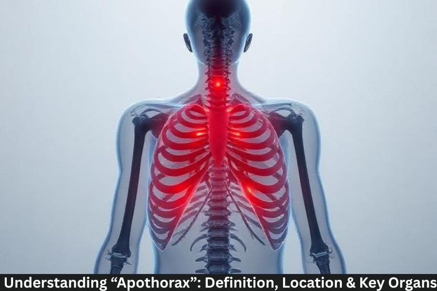
What Is the Apothorax?
Simple Definition
The apothorax refers to the lower portion of the thorax, located just above the diaphragm. It acts as a transitional zone between the chest cavity and the upper abdominal region.
Why the Term Is Not Common in Modern Textbooks
Modern anatomy generally uses broader, clearer terms such as “thoracic cavity.” The apothorax is mainly mentioned in classical anatomy and comparative anatomy.
Anatomical Location of the Apothorax
Lower Thoracic Region
The apothorax lies in the lower chest area, mostly around ribs 8–12.
Borders of the Apothorax
- Upper boundary: Middle thorax
- Lower boundary: Diaphragm
- Side boundaries: Lower ribs and costal cartilage
Relation to the Diaphragm
The diaphragm forms the floor of the apothorax, influencing its movement during breathing.
Structure of the Apothorax
Ribs and Vertebrae Involved
The apothorax is supported by:
- Ribs 8–12
- Thoracic vertebrae T8–T12
- Costal cartilages
Muscles in the Region
Key muscles include:
- Intercostal muscles
- Lower thoracic muscles
- Diaphragmatic attachments
Soft Tissues and Connective Support
The region contains:
- Fascia
- Intercostal spaces
- Nerves and blood vessels
Key Organs in the Apothorax
Lower Lobes of the Lungs
The majority of the lower lung lobes fall within this region.
Base of the Heart
The lower part of the heart rests close to the apothorax.
Esophagus and Major Vessels
Important structures passing through include:
- Esophagus
- Aorta
- Inferior vena cava (IVC)
Functions of the Apothorax
Role in Breathing
The apothorax expands and contracts during diaphragm movement, helping the lungs fill with air.
Protection of Vital Organs
It shields major organs, including parts of:
- Heart
- Lungs
- Blood vessels
Support in Body Movements
It stabilizes the upper body during bending, twisting, and lifting.
Apothorax vs. Thorax
Major Differences
- The thorax includes the entire chest region.
- The apothorax is the lower segment of the thorax.
How the Apothorax Fits into the Thoracic Cavity
It supports lower respiratory movement and forms a boundary with the abdominal cavity.
Comparison Table
| Feature | Thorax | Apothorax |
|---|---|---|
| Area | Entire chest cavity | Lower thoracic region |
| Ribs Involved | 1–12 | Mostly 8–12 |
| Key Organs | Heart, lungs, vessels | Lower lungs, heart base |
| Function | Protection + breathing | Lower thoracic support |
Clinical Significance
Conditions That Affect This Region
- Lower rib fractures
- Diaphragm spasms
- Lung infections
- Chest trauma
Importance in Respiratory Examinations
Doctors observe:
- Lower lung expansion
- Diaphragm movement
- Fluid accumulation in lower lungs
Conclusion
The apothorax may not be a widely used anatomical term today, but understanding it helps students visualize how the lower thoracic structures work together. Knowing its position, boundaries, and associated organs strengthens your knowledge of respiratory anatomy and overall thoracic function. Whether you’re preparing for an exam or revising key concepts, the apothorax is an essential structure worth understanding.
FAQs
1. What is the apothorax?
It is the lower part of the thorax located just above the diaphragm.
2. Is the apothorax a separate cavity?
No, it is a regional subdivision of the thorax.
3. What organs are found in the apothorax?
Lower lung lobes, base of the heart, esophagus, and major vessels.
4. Why is the apothorax important?
It plays key roles in breathing, organ protection, and body support.
5. Do modern textbooks mention the apothorax?
Not frequently—it’s more common in older anatomical descriptions.